lunes, 26 de noviembre de 2012

Nuevas pistas sobre el origen del autismo abre el camino a nuevos tratamientos


Tras identificar una síntesis particular de una determinada proteína, científicos de Canadá pudieron revertir algunos síntomas en ratones. Según los expertos, se abre el camino hacia nuevas terapéuticas, incluso farmacológicas.
La noticia se publicó en Nature y está dando la vuelta al mundo: la evidencia directa de que una alteración en la síntesis de proteínas puede provocar comportamientos autistas en ratones abre una posible vía para identificar nuevas dianas terapéuticas para los síndrome relacionados con el espectro autista.

Los investigadores de la Universidad McGill y la Universidad de Montreal (Canadá) aseguran haber identificado un vínculo crucial entre la síntesis de proteínas y los trastornos del espectro autista, un hallazgo que, a su juicio, puede impulsar el desarrollo nuevas vías terapéuticas.

La regulación de la síntesis de proteínas, también conocida como ARNm, es el proceso por el cual las células fabrican proteínas. Este mecanismo, explican los investigadores, está involucrado en todos los aspectos de la función celular y del organismo. Los investigadores encontraron que la síntesis anormalmente elevada de un grupo de proteínas neuronales, llamadas neuroliginas, causa síntomas similares a los que se diagnostica en los casos de trastornos del espectro autista.

El estudio reveló que las conductas asociadas al autismo pueden ser rectificadas en los ratones adultos con compuestos que inhiben la síntesis de proteína, o con una terapia génica que tenga como diana la neuroliginas.

El equipo estaba investigando, en realidad, el papel de la síntesis de proteínas en la etiología del cáncer, y vieron con sorpresa que mecanismos similares a los que investigan pueden estar asociados en el desarrollo del autismo.

Según explica el profesor Nahum Sonenberg, del Departamento de Bioquímica de McGill, Facultad de Medicina y el Centro de Investigación del Cáncer Goodman, emplearon un modelo de ratón en el que se eliminó un gen clave que controla el inicio de la síntesis de proteínas. "Vimos que se incrementaba la producción de neuroliginas, proteínas importantes en la formación y regulación de las conexiones conocidas como sinapsis entre las células neuronales en el cerebro y esenciales para el mantenimiento del equilibrio en la transmisión de información de una neurona a otra".

El trabajo, subrayan, es el primero en relacionar la traducción de control de neuroliginas con la alteración de la función sináptica y los comportamientos similares al autismo en ratones.

Neurodesarrollo
Los trastornos del espectro autista abarcan una amplia gama de enfermedades del neurodesarrollo que afectan a tres áreas de comportamiento: interacción social, comunicación y comportamientos e intereses restringidos, repetitivos y estereotipados.
Se calcula que entre 4 y 20 de cada 10.000 niños de la población general padecen un trastorno del espectro autista. Generalmente es cuatro a cinco veces más frecuente en niños que en niñas, y no se asocia con ningún grupo socioeconómico. Generalmente se detecta en los primeros 2 o 3 años de la vida del niño.

Fuente: Nature

domingo, 11 de noviembre de 2012

Nuevos avances sobre el tinnitus

Las investigaciones acerca del tinnitus o acúfenos han proporcionado importantes hallazgos durante el último año sobre el tinnitus.

Se ha descubierto que el riesgo de padecer tinnitus o acúfenos es mayor bajo estrés. 
Otro estudio demuestra que el tinnitus lo produce el propio cerebro para compensar la existencia de una pérdida de audición. 
En cuanto a los tratamientos, la terapia que se basa en la aceptación y la conciencia plena ayuda al paciente a convivir con el tinnitus. 
Se ha demostrado también que la terapia a través de internet es prácticamente igual de efectiva que las terapias de grupo presenciales. 
Asimismo, la terapia de reinicio acústico coordinado ayuda a reducir la intensidad y las molestias del tinnitus. Por último, un estudio británico pone de manifiesto que cuatro de cada diez personas encuestadas desconocen lo que es el tinnitus.

La incidencia de tinnitus se duplica bajo estrés

Los expertos creen que existe una relación directa entre el tinnitus y el estrés. La incidencia de los casos de tinnitus se duplica en personas que sufren estrés, según indica un estudio realizado por el Instituto Karolinska en Suecia.

“Descubrimos que el tinnitus afectaba 2,5 veces más a personas que estaban en situaciones de estrés prolongadas”, explica el Profesor Barbera Canlon, investigador jefe del estudio.

Más que un problema de audición

Los investigadores han descubierto que el cerebro es el responsable del pitido de oídos que produce el tinnitus, como estrategia para compensar la existencia de una pérdida de audición.
Según el estudio realizado en el Centro Médico de la Universidad de Georgetown, en Estados Unidos, el tinnitus no se produce solo por un efecto oclusivo o daño en el oído, sino que es el resultado fallido del propio cerebro por recuperarse.

Elegir la terapia apropiada

Según una tesis doctoral en psicología presentada en Suecia, la terapia basada en la aceptación y la conciencia plena puede servir para aprender a convivir con el tinnitus. Esta técnica busca generar una nueva actitud hacia el tinnitus, para evitar que los síntomas consuman la vida del paciente. Este método se denomina Terapia de Aceptación y Compromiso.

Terapia a través de internet

Investigadores de la Universidad Johannes Gutenberg en Alemania y de la Universidad de Linköping en Suecia trataron a un grupo de pacientes con tinnitus de moderado a severo con distintos tipos de terapias, durante un periodo de diez semanas. Los resultados del estudio ponen de manifiesto que la terapia a través de internet es igual de efectiva que la terapia de grupo presencial.
Al comparar los resultados con el grupo de control, cuyos sujetos solo participaron en un foro online, se observó que los pacientes que siguieron la terapia a través de internet o participaron en la terapia de grupo podían controlar mejor los síntomas del molesto sonido del tinnitus que perciben en sus oídos.

Terapia de reinicio acústico coordinado

Un ensayo clínico ha demostrado que una terapia denominada neuromodulación de reinicio acústico coordinado (Acoustic Coordinated Reset, en inglés), reduce la intensidad y el malestar que produce el tinnitus en siete de cada diez pacientes. Este estudio comparaba los resultados de la terapia de reinicio acústico coordinado con un tratamiento placebo de control en 63 pacientes con tinnitus de larga duración.

En el ensayo clínico, los pacientes tenían que llevar unos auriculares especiales durante varias horas al día. Los auriculares emitían una serie de tonos ajustados de acuerdo a la frecuencia característica del tinnitus del paciente. De esa forma, se pretendía interrumpir los patrones rítmicos del ruido del tinnitus que producen las células del nervio auditivo del cerebro.

Cuatro de cada diez desconocen lo que es el tinnitus

Por último, un estudio realizado por la organización británica de discapacitados auditivos “Action on Hearing loss” (Acción ante la Pérdida de Audición), revela que el 39% de los encuestados desconocían lo que era el tinnitus o acúfenos, y el 22% pensaba incluso que consistía en tener alergia al metal.
Para mas informacion sigua estos links
El tinnitus afecta hasta 2.5 veces mas a personas con estres
Tinnitus: Mas que una problema de audicion
Hacer frente al tinnitus depende de la terapia adecuada
La terapia para el tinnitus a traves de internet resulta util
La terapiad de reinicio acustico coordinado (ACR) ayuda a reducir los sintomas del tinnitus
Cuatro de cada diez desconocen lo que es el tinnitus


Fuente:  http://www.spanish.hear-it.org/Nuevos-avances-sobre-el-tinnitus
Julio de 2012

The Worst Noises in the World: Why We Recoil at Unpleasant Sounds

ScienceDaily (Oct. 12, 2012) — Heightened activity between the emotional and auditory parts of the brain explains why the sound of chalk on a blackboard or a knife on a bottle is so unpleasant.

 ScienceDaily (12 de octubre de 2012) - Una mayor actividad entre las partes emocionales y auditivas del cerebro explica por qué el sonido de la tiza en una pizarra o un cuchillo en una botella es tan desagradable.


In a study published today in the Journal of Neuroscience and funded by the Wellcome Trust, Newcastle University scientists reveal the interaction between the region of the brain that processes sound, the auditory cortex, and the amygdala, which is active in the processing of negative emotions when we hear unpleasant sounds.

Brain imaging has shown that when we hear an unpleasant noise the amygdala modulates the response of the auditory cortex heightening activity and provoking our negative reaction.

"It appears there is something very primitive kicking in," says Dr Sukhbinder Kumar, the paper's author from Newcastle University. "It's a possible distress signal from the amygdala to the auditory cortex."

Researchers at the Wellcome Trust Centre for Neuroimaging at UCL and Newcastle University used functional magnetic resonance imaging (fMRI) to examine how the brains of 13 volunteers responded to a range of sounds. 

Listening to the noises inside the scanner they rated them from the most unpleasant -- the sound of knife on a bottle -- to pleasing -- bubbling water. 

Researchers were then able to study the brain response to each type of sound.

Researchers found that the activity of the amygdala and the auditory cortex varied in direct relation to the ratings of perceived unpleasantness given by the subjects. 

The emotional part of the brain, the amygdala, in effect takes charge and modulates the activity of the auditory part of the brain so that our perception of a highly unpleasant sound, such as a knife on a bottle, is heightened as compared to a soothing sound, such as bubbling water.

Analysis of the acoustic features of the sounds found that anything in the frequency range of around 2,000 to 5,000 Hz was found to be unpleasant. 

Dr Kumar explains: "This is the frequency range where our ears are most sensitive. 

Although there's still much debate as to why our ears are most sensitive in this range, it does include sounds of screams which we find intrinsically unpleasant."

Scientifically, a better understanding of the brain's reaction to noise could help our understanding of medical conditions where people have a decreased sound tolerance such as hyperacusis, misophonia (literally a "hatred of sound") and autism when there is sensitivity to noise.
Professor Tim Griffiths from Newcastle University, who led the study, says: "This work sheds new light on the interaction of the amygdala and the auditory cortex. 

This might be a new inroad into emotional disorders and disorders like tinnitus and migraine in which there seems to be heightened perception of the unpleasant aspects of sounds."

Most Unpleasant Sounds

Rating 74 sounds, people found the most unpleasant noises to be:
  1. Knife on a bottle
  2. Fork on a glass
  3. Chalk on a blackboard
  4. Ruler on a bottle
  5. Nails on a blackboard
  6. Female scream
  7. Anglegrinder
  8. Brakes on a cycle squealing
  9. Baby crying
  10. Electric drill
Least Unpleasant Sounds

  • Applause
  • Baby laughing
  • Thunder
  • Water flowing
 
Fuente:  
http://www.sciencedaily.com

Medical Devices Powered by the Ear Itself

ScienceDaily (Nov. 8, 2012) — 
For the first time, researchers power an implantable electronic device using an electrical potential -- a natural battery -- deep in the inner ear.

Por primera vez, los investigadores alimentan un dispositivo implantable electrónico utilizando un potencial eléctrico - una batería natural - profunda  del propio oído interno.


The chip is small enough to fit in the cavity of the middle ear. (Credit: Patrick P. Mercier)


Deep in the inner ear of mammals is a natural battery -- a chamber filled with ions that produces an electrical potential to drive neural signals. 
In a recent issue of the journal Nature Biotechnology, a team of researchers from MIT, the Massachusetts Eye and Ear Infirmary (MEEI) and the Harvard-MIT Division of Health Sciences and Technology (HST) demonstrate for the first time that this battery could power implantable electronic devices without impairing hearing.
The devices could monitor biological activity in the ears of people with hearing or balance impairments, or responses to therapies.

Eventually, they might even deliver therapies themselves.
In experiments, Konstantina Stankovic, an otologic surgeon at MEEI, and HST graduate student Andrew Lysaght implanted electrodes in the biological batteries in guinea pigs' ears. 

Attached to the electrodes were low-power electronic devices developed by MIT's Microsystems Technology Laboratories (MTL). 

After the implantation, the guinea pigs responded normally to hearing tests, and the devices were able to wirelessly transmit data about the chemical conditions of the ear to an external receiver.

"In the past, people have thought that the space where the high potential is located is inaccessible for implantable devices, because potentially it's very dangerous if you encroach on it," Stankovic says.

 "We have known for 60 years that this battery exists and that it's really important for normal hearing, but nobody has attempted to use this battery to power useful electronics."

The ear converts a mechanical force -- the vibration of the eardrum -- into an electrochemical signal that can be processed by the brain; the biological battery is the source of that signal's current. 

Located in the part of the ear called the cochlea, the battery chamber is divided by a membrane, some of whose cells are specialized to pump ions. 

An imbalance of potassium and sodium ions on opposite sides of the membrane, together with the particular arrangement of the pumps, creates an electrical voltage.

Although the voltage is the highest in the body (outside of individual cells, at least), it's still very low. 

Moreover, in order not to disrupt hearing, a device powered by the biological battery can harvest only a small fraction of its power.

Low-power chips, however, are precisely the area of expertise of Anantha Chandrakasan's group at MTL.
The MTL researchers -- Chandrakasan, who heads MIT's Department of Electrical Engineering and Computer Science; his former graduate student Patrick Mercier, who's now an assistant professor at the University of California at San Diego; and Saurav Bandyopadhyay, a graduate student in Chandrakasan's group -- equipped their chip with an ultralow-power radio transmitter: After all, an implantable medical monitor wouldn't be much use if there were no way to retrieve its measurements.

But while the radio is much more efficient than those found in cellphones, it still couldn't run directly on the biological battery. 

So the MTL chip also includes power-conversion circuitry -- like that in the boxy converters at the ends of many electronic devices' power cables -- that gradually builds up charge in a capacitor. 

The voltage of the biological battery fluctuates, but it would take the control circuit somewhere between 40 seconds and four minutes to amass enough charge to power the radio. 

The frequency of the signal was thus itself an indication of the electrochemical properties of the inner ear.


To reduce its power consumption, the control circuit had to be drastically simplified, but like the radio, it still required a higher voltage than the biological battery could provide. 

Once the control circuit was up and running, it could drive itself; the problem was getting it up and running.


The MTL researchers solve that problem with a one-time burst of radio waves. "In the very beginning, we need to kick-start it," Chandrakasan says. "Once we do that, we can be self-sustaining. The control runs off the output."

Stankovic, who still maintains an affiliation with HST, and Lysaght implanted electrodes attached to the MTL chip on both sides of the membrane in the biological battery of each guinea pig's ear. 

In the experiments, the chip itself remained outside the guinea pig's body, but it's small enough to nestle in the cavity of the middle ear.
Cliff Megerian, chairman of the otolaryngology department at Case Western Reserve University, says that he sees three possible applications of the researchers' work: in cochlear implants, diagnostics and implantable hearing aids. 

"The fact that you can generate the power for a low voltage from the cochlea itself raises the possibility of using that as a power source to drive a cochlear implant," Megerian says. 

"Imagine if we were able to measure that voltage in various disease states. 

There would potentially be a diagnostic algorithm for aberrations in that electrical output."
"I'm not ready to say that the present iteration of this technology is ready," Megerian cautions. 

But he adds that, "If we could tap into the natural power source of the cochlea, it could potentially be a driver behind the amplification technology of the future."

The work was funded in part by the Focus Center Research Program, the National Institute on Deafness and Other Communication Disorders, and the Bertarelli Foundation.

Key words: electrical, potential, inner ear, deep,journal Nature Biotechnology,MIT,Konstantina, Stankovic, Andrew, Lysaght,Anantha Chandrakasan, Patrik Mercier, Saurav Bandyopadhyay,Cliff Megerian,Case Western Reserve University,Focus Center Research Program, the National Institute on Deafness and Other Communication Disorders, and the Bertarelli Foundation.

Fuente:  
http://www.sciencedaily.com/releases/2012/11/121108151730.htm

sábado, 10 de noviembre de 2012

Hearing Health Summit

BP AndreaBoidman_L

"The Promise of Cell Regeneration"


On October 3, 2011, Bryan Pollard represented Hyperacusis Research at the Summit on Hearing Restoration at the New York Academy of Medicine. 

This Summit was sponsored by the Hearing Health Foundation (formerly Deafness Research Foundation). It brought together the leaders in the field of cell regeneration research to discuss what needs to be accomplished for therapy for cell regeneration in the ear to become a reality. 
Learn more about Hearing Health Foundation's ground-breaking Hearing Restoration Project. 
While this Summit did not relate directly to issues with Hyperacusis, the Hearing Restoration Project is an important effort because hair cell damage is still a possible consideration in the etiology of Hyperacusis. 
Also Hyperacusis Research is working together to sponsor a future research project with the Hearing Health Foundation.


Panel Discussion

HearingHealth Panel_L 


The event started out with a panel discussion lead by Dr. Jon Lapook of the CBS Evening News. 

The members of the panel were: Dr. Sujana Chandrasekhar from New York Otology, Dr. Ed Rubel from the University of Washington, Dr. Stefan Heller from Stanford University and Dr. Andy Groves from Baylor College of Medicine. 

 An interesting perspective from the panel discussion was how much less the NIH funds hearing research compared to cancer and other significant health issues. 

That was an important insight into possibly why it is even more difficult to get funding for hyperacusis since it represents only a fraction of the population with ear issues. 

Dr. Lopook referenced a recent news story highlighting the growing risk of hearing loss for children. There was a good question about why there were not more news stories on hearing challenges. 
Dr. Lopook described the significant challenge of getting even one deeper story of 5 or 6 minutes into the evening news every few weeks.
Dr. Chandrasekhar gave a good overview the basic clinical types of cases she works with. She supports efforts with veterans who have hearing damage from the battle field.

Dr. Rubel described the historical work that was accomplished to discover that birds and reptiles re-grow hair cells after damage. 

This gives a great model to study to understand why mammals are different since no mammal can re-grow hair cells after damage. 

Dr Heller gave a short description of how various types of stem cells can be used as a basis to initiate new hair cell growth. Finally Dr. Groves described briefly how hair cells grow. Each of these researchers gave a much more detailed description in their presentation.

How We Hear

The first speaker was Dr. George Gates who is on the board of the Hearing Health Foundation. His presentation gave a good overview of the basic ear function including the inner ear and hair cell function.

He described how the outer hair cells (OHC) work from feedback signals to amplify quiet sounds. I asked a follow-up question about what other function the OHC has and he responded by describing that the OHC does provide some function to dampen loud sounds.

Hair Cell Regeneration

Dr. Rubel's presentation gave more details on birds and reptiles amazing hair cell recovery and re-growth from damage. 

For example, birds re-grow their hair cells in 22 days. His session ended with a question on Tinnitus. Another question related to both Tinnitus and Hyperacusis where a member of the audience referenced work in Germany to use lower level laser treatments to improve the condition of Tinnitus and Hyperacusis patients. No one on the panel was familiar with the work.

Barriers to Hair Cell Regeneration

Dr. Andy Groves presented details on hair cell regeneration that were highly technical. It appears there is a gene (P27) which won't allow hair cells to divide once cells are older. 

 A key element to overcome this issue relates to Notch Signaling where one cell gives signals to the cell next to it which prevent the cell from changing into a different type of cell. Suffice it to say that there are some serious barriers to be overcome.

Stem Cells and Inner Ear Cell Regeneration

Dr. Heller described how stem cells work. He reviewed the 3 types of stem cells which enables various methods to obtain the needed building blocks. 

He then described how the process works to move from a stem cell to grow a specific type of cell such as a hair cell. 

This was also very technical and while occasional media stories make this sound like it is ready for prime time for certain diseases there are still huge barriers to overcome.

Conclusion

BP Speak_Spon_LThe key take-away is that while barriers remain large, the team felt overall that a real pathway is possible in the next decade or two. 

The most important aspect that relates to hyperacusis is that this research work will continue to drive the need for more detailed understanding of every inner ear component down to the molecular level. 

Every new piece of knowledge gained may further advance possible models relating to hyperacusis as long as we are connected to the work. 

For example, a key issue with hyperacusis is getting solid evidence for whether or not hyperacusis patients have hair cell damage. 

Currently all methods to truly establish hair cell damage can only be done on a destroyed cochlea. In a follow-up discussion, 

Dr. Heller described that recent advances in optical tomography may soon enable this diagnostic tool to be able to view inside a live cochlea. 

That breakthrough would enable many in the study of the inner ear to gain much needed understanding for modeling the inner ear and help to finally answer the question for damage for hyperacusis patients.

fuente: Hyperacusis Research

Product Experiences

SoundMeterIPhone.133180935_std
There are inexpensive consumer level sound meters such as the Scosche - SPL Meter that can give you the sound dB level of any product. Also, there are now app's to add to a smart phone to enable any phone to provide sound dB levels – although this will tend to be less accurate than a dedicated meter. Click here for an iphone sound meter app. Since consumer level sound meters don't provide a way to test frequency, hearing from the experiences of others is a helpful way to make better choices. Please share your product experiences in the comment section below. Please include sound dB levels if you have measured it or if the manufacturer provides a level based on their tests.


Fuente: http://hyperacusisresearch.org/index.php?option=com_content&view=category&layout=blog&id=13&Itemid=64

Nota, hay un video en youtube de este equipo

 <iframe width="640" height="360" src="http://www.youtube.com/embed/JGAFmAALjJQ" frameborder="0" allowfullscreen></iframe>

 fuente:http://www.youtube.com/watch?v=JGAFmAALjJQ

Hyperacusis Research

cochlea
The list of possibilities for hyperacusis mechanisms is still quite large. 

To narrow the discussion, we focus primarily on cases induced by acoustic trauma.

It is natural then to start with the middle ear function for possible models. 

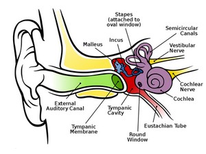
The stapedius muscle is responsible for sound attenuation by dampening the vibration of the stapes. 

Since many hyperacusis patients appear to have a normal stapedial reflex test (by current testing methods), there are very few works on the topic. 

In a work on the "Functional Model of the Middle Ear Ossicles," the authors suggest a mechanism of how paralysis of the stapedius muscle, caused by an injury to the facial nerve, results in hyperacusis

Further insights might be gained by a deeper analysis and possible advancement of the various acoustic reflex testing methods comparing a hyperacusis population to a control group.

Many possible mechanisms for hyperacusis start in the cochlea. Baguley and Andersson's book references Sahley and Nodar's 2001 article on "A Biochemical Model of Peripheral Tinnitus." 

A summary from their abstract states: "Naturally occurring opioid dynorphins are released from lateral efferent axons into the synaptic region beneath the cochlear inner hair cells during stressful episodes. 

In the presence of dynorphins, the excitatory neurotransmitter glutamate, released by inner hair cells in response to stimuli or (spontaneously) in silence, is enhanced at cochlear NMDA receptors. 

This results in altered neural excitability and/or an altered discharge spectrum in (modiolar-oriented) type I neurons normally characterized by low rates of spontaneous discharge and relatively poor thresholds. 

It is also possible that chronic exposure to dynorphins leads to auditory neural excitotoxicity via the same receptor mechanism." 

They propose this spontaneous activity leads to the perception of tinnitus and that the perceived intensity of sound may increase resulting in hyperacusis

This work is an example of where model associations between tinnitus and hyperacusis can be difficult. 

It would be far better to have an analysis of similar causal mechanisms purely in relationship to hyperacusis.

There are a wide range of supporting hyperacusis works describing the connections to the cochlea. 

The case of a musician who had his cochlea destroyed in a mostly deaf ear to relieve his symptoms is interesting as later works suggest such a radical measure may not impact the problem.

Some works suggest an abnormality of outer hair cell (OHC) function. 

In a work entitled "DPOAE in Tinnitus Patients with Cochlear Hearing Loss considering Hyperacusis and Misophonia," the authors promote the model that the OHC's are the most probable place generating tinnitus. 

They find that decreasing of otoemission "DPOAE amplitudes in patients with cochlear hearing loss and tinnitus suggests significant role of OHC pathology, unbalanced by IHC injury in generation of tinnitus in patients with hearing loss of cochlear localization.


DPOAE fine structure provides us the additional information about DPOAE amplitude recorded in two points per octave, spreading the amount of frequencies f2, where differences are noticed in comparison of two groups - tinnitus patients and control.

Function growth rate cannot be the only parameter in estimation of DPOAE in tinnitus patients with cochlear hearing loss, also including subjects with hyperacusis and misophony. 

Hyperacusis has important influence on DPOAE amplitude, increases essentially amplitude of DPOAE in the examined group of tinnitus patients.

" Another work focused on the OHC's impact from 1996 is entitled "Nonlinearity of mechanoelectrical transduction of outer hair cells as the source of nonlinear basilar-membrane motion and loudness recruitment." 

According to the experiment's data and "the feedback concept of outer hair cell action, disruption of the mechanoelectrical transduction of OHC leads to both a reduction of gain and linearizing of the response; that is, to both hearing loss and loudness recruitment.

" Since recruitment and hyperacusis are quite different, this model should not be extented to hyperacusis

However, since most OHC works were not primarily focused on hyperacusis, it may be useful to have a dedicated project that can conclusively prove OHC function in typical hyperacusis patients.


Dr. J. A. Vernon at the Oregon Health and Science University provided a basic clinical overview in 2002 entitled "Hyperacusis: Testing, Treatments, and a Possible Mechanism." He describes background information on the topic and the pink noise treatment program he has used for patients. 

He concludes with a brief summary of the model often accepted for the cause: "It appears that the normal function of the olivocochlear bundle, which supplies efferent innervation to the cochlea, is to exert a suppressive or inhibitor effect upon responses to incoming sounds. 

If these efferent nerves are not performing normally it seems somewhat reasonable to propose that all sounds might be perceived as being louder than usual."

One classic work that led to moving beyond the cochlea is a paper from 2000 by Salvi, Wang, and Ding at University of Buffalo entitled: "Auditory plasticity and hyperactivity following cochlear damage." 

In it they describe some of the "functional changes that occur in the central auditory pathway after the cochlea is damaged by acoustic overstimulation or by carboplatin, an ototoxic drug that selectively destroys inner hair cells (IHCs). 

Acoustic trauma typically impairs the sensitivity and tuning of auditory nerve fibers and reduces the neural output of the cochlea.

Surprisingly, our results show that restricted cochlear damage enhances neural activity in the central auditory pathway. 

Despite a reduction in the auditory-nerve compound action potential (CAP), the local field potential from the inferior colliculus (IC) increases at a faster than normal rate and its maximum amplitude is enhanced at frequencies below the region of hearing loss. 

To determine if this enhancement was due to loss of sideband inhibition, we recorded from single neurons in the IC and dorsal cochlear nucleus before and after presenting a traumatizing above the unit's characteristic frequency (CF). 

Following the exposure, some neurons showed substantial broadening of tuning below CF, less inhibition, and a significant increase in discharge rate, consistent with a model involving loss of sideband inhibition. 

Selective IHC loss reduces the amplitude of the CAP without affecting the threshold and tuning of the remaining auditory nerve fibers. 

Although the output of the cochlea is reduced in proportion to the amount of IHC loss, the IC response shows only a modest amplitude reduction, and remarkably, the response of the auditory cortex is enhanced. 

These results suggest that the gain of the central auditory pathway can be up- or down regulated to compensate for the amount of neural activity from the cochlea." 

Although this work is centered on hearing loss, there are some important details which may relate to hyperacusis

 Figure 3 C from their initial experiment where chinchillas were exposed to 105db for 5 days shows the overshoot of the IC amplitude just above the 60dB level which exceeds the pre-exposure levels by a significant amount.


In another experiment where the IHC's only were selectively destroyed resulting in a similar reduction in output of the cochlea (lower CAP), the authors also find an increase in AC amplitude. 

Thus they propose that a mechanism for tinnitus could be "the loss of lateral inhibition that unmasks pre-existing neural circuits within the auditory pathway." 

Additionally they state that since "neural activity is enhanced and that amplitude grows at an abnormally rapid rate following cochlear damage, it seems plausible that recruitment and hyperacusis originate in the central auditory pathway.}

 While the model seems to fit well for recruitment, it does not fit as well for typical hyperacusis patients since they often have normal audiograms and do not have hearing loss in the lower thresholds. 

Additionally, the work is entirely based on a scenario created by hair cell damage (either IHC and OHC or both) which further supports the need to establish if typical hyperacusis patients have hair cell damage.


In their book (latest version from 2008) entitled Tinnitus Retraining Therapy, Dr. Jastreboff and Dr. Hazell devote the second chapter to "The neurophysiological model of tinnitus and decreased sound tolerance.

" Their primary theory is called the "discordant dysfunction (damage) theory." They postulate that "the tinnitus signal originates in the inner ear when one type of sensory cell, OHC, is more dysfunctional than the other type of sensory cells, IHC, at the same area of the basilar membrane in the cochlea. 

When neurons in the dorsal cochlear nuclei receive excitation from IHC but not from the damaged OHC, then an imbalance occurs at this level of the auditory system. 

This, in turn, causes abnormal activity in the form of burst of high-frequency neuronal discharges, which, after amplification within the auditory system are perceived as tinnitus." 

It is interesting to contrast the theory here focused on OHC loss compared to Salvi's work that was more focused on IHC loss. 

On the issue of detectable hearing loss for tinnitus, they state that "20% of patients with tinnitus have normal hearing. 

This is because changes are too small to be detectable on a standard audiogram." 

A key point to note here is that there is a wide frequency range above the normal 8000Hz range of standard audiograms that could be explored to help confirm these possibilities. In moving from the model above on tinnitus to the model associated with hyperacusis, Jastreboff is less detailed. 

The book simply states that "the model predicts that a relatively high percentage of tinnitus patients should exhibit increased sensitivity to external sound, that is decreased sound tolerance. 

This is what one would expect from the increased auditory gain, or the combination of an increased gain and some even minor dysfunction of the inner ear.

" Later it is further explained that in hyperacusis, "the external signal undergoes abnormal enhancement/amplification within the subconscious auditory pathways and only secondarily activates the limbic and autonomic nervous systems.

" The opportunity for further works here is similar to above models: prove the functional capabilities of the OHC's in hyperacusis patients.
In 2003, Formby, Sherlock, and Gold presented a paper focused on proving the central gain control process entitled: "Adaptive plasticity of loudness induced by chronic attenuation and enhancement of the acoustic background."

In it the authors show that "after continuous, 2-week earplugging and low-level noise treatments that listeners become more and less sensitive, respectively, to the loudness of sounds. 

This simple demonstration of adaptive plasticity is consistent with modification of a theoretical gain control process, which is the basis for desensitizing sound therapies used in treating hyperacusis and related sound tolerance problems." 

The specific mechanism of adaptive plasticity is not proposed. While the loudness judgments using the Contour Test of Loudness Perception in the work is helpful, another key component for direct comparison to hyperacusis would have been actual levels and types of pain the participants experienced when exposed back to everyday loud noises. 

More specifically did the participants experience pain for the days following the experiment.

Additional opportunities for research relate to a more detailed assessment of hyperacusis patients' symptoms. 

Most surveys are trying to establish whether or not a person has hyperacusis

Baugley has summarized some of these prevalence surveys which show a wide range of results from 8-9% in a Swedish study to 15% in a Polish study. 

He also summarizes the following finding: "Among patients attending tinnitus clinics with a primary complaint of tinnitus the prevalence of hyperacusis is about 40%; and in patients with a primary complaint of hyperacusis the prevalence of tinnitus has been reported as 86%". 

Beyond the issue of lack of standardization in questionnaires (one by Khalfa is presented as a possible standard), the more important detail for researches is a detailed guide of symptoms beyond life impacts. 

For example, with many neuropathies the Leeds Assessment of Neuropathic Symptoms and Signs (LANSS) Pain Scale is used. 

Similarly, in hyperacusis it would be helpful to know the level of pain severity, the type of pain such as throbbing, constant, random, the location of pain, the length of pain and timing after excessive noise exposure. 

This may yield possible measurable impacts. These results may add insights to psychological mechanisms and models associated with hyperacusis.

Fuente: http://hyperacusisresearch.org/index.php?option=com_content&view=article&id=16:hearing-protection&catid=5:research&Itemid=71

Hyperacusis Workshop at 2013 ARO Midwinter Meeting


aro 2013 


Hyperacusis Research is pleased to announce that the 2013 Association for Research in Otolaryngology (ARO)  midwinter meeting will include a workshop on Hyperacusis.


This effort was made possible by a partnership with Dr. Peter Steyger from the Oregon Hearing Research Center at OHSU (Oregon Health and Science University) and the Hearing Health Foundation.  

The ARO is an international scientific society composed of basic scientists and clinicians who are actively investigating basic science and clinical problems associated with hearing, speech, balance, smell, taste and diseases of the head and neck. 

The ARO provides a forum for the interchange of information through its peer reviewed journal, JARO, and via a professional yearly meeting.  

We are excited to have this audience exposed the challenges of hyperacusis.

Fuente: http://hyperacusisresearch.org/

Beca para Revisión de la Literatura sobre hiperacusia

HHF Research Hyperacusis Research se complace en anunciar su primera donación para  investigación.  
Esta donación prestará apoyo a la donación de la Hearing Health Foundation para la investigación sobre una  revisión de la literatura centrada en la hiperacusia, reclutamiento, misophonia, fonofobia y mecanismos de volumen / intensidad del procesamiento en la vía auditiva.
WomanEarHand4Hay varios objetivos clave de la búsqueda en la literatura. En primer lugar, el objetivo es proporcionar definiciones para cada trastorno (teniendo en cuenta que puede haber definiciones múltiples para una sola enfermedad) y si existe una prevalencia entre los roles de las definiciones específicas.

A continuación, se describira un desarrollo global para cada trastorno y  dar ejemplos de cada condición. Por último, el documento ofrecerá una propuesta de mecanismo o etiología demostrada, si esta fuera posible.
La
Hearing Health Foundation ha seleccionado al beneficiario de las subvenciones de  revisión de la literatura .  

El investigador principal será el Dr. Richard S. Tyler de la Universidad de Iowa, Departamento de Patología del Habla y Audiología.(Brian C. J. Moore será el co-autor.) 

Estamos muy entusiasmados con esta elección dado que el Dr. Tyler y su departamento en la Universidad de Iowa tienen un excelente historial de 30 años en investigación del tinnitus y la hiperacusia .  
Él fue el primero en mostrar que los pacientes con tinnitus a menudo tienen hiperacusia.  
Anteriormente ha escrito artículos de revisión que incluyeron la hiperacusia como tema principal. Para más información sobre el Dr. Tyler ver su sitio web en la Universidad de Iowa.
¿Por qué hacer una revisión de la literatura?
Para los investigadores, este esfuerzo de revisión de la literatura es un paso vital inicial al intentar mover un esfuerzo de investigación que se ha enfrentado a retos importantes en la toma de grandes descubrimientos. Para los que sufren de hiperacusia, este paso puede parecer innecesario, especialmente si usted ha hecho su propia revisión de la literatura. 

  
Sin embargo, el tiempo dedicado a la obtención de una puesta al día resumida y publicada será muy importante para asegurar l que los esfuerzos próximas  se encaminan en la dirección correcta y no redundante que cubre otros temas que ya se han investigado. También se asocia con la Fundación  a los desafíos que enfrenta la investigación hiperacusia se comunicará a una base mucho más amplia de investigadores del oído.

RESEARCH Literature review research opportunity focused on hyperacusis, recruitment, misophonia, phonophobia, and mechanisms
GRANT OPPORTUNITY


ELIGIBILITY Hearing Health Foundation (HHF), formerly Deafness Research Foundation (DRF), is the leading national source of private funding for research in hearing and balance science. HHF requests applications from junior and senior-level investigators to research and catalog existing, recent (within the last 25 years) literature on hyperacusis, misophonia, phonophobia, recruitment, and mechanisms of volume/intensity processing in the auditory pathway.


REQUIREMENTS /OBJECTIVES
Literature search will be conducted using pubmed and other biomedical or audiological indices, and the original literature obtained. All statements and conclusions will be referenced/cited to the original literature. Reviews and speculative hypotheses will not be sufficient.
Finished report must (a) provide definitions discovered during literature review for each disorder (noting that there may be multiple definitions for a single disorder) and whether there is a prevalence among papers for specific definitions (b) provide comprehensive background to each disorder (as possible); (c) give examples of each condition; and (d) provide a proposed mechanism or demonstrated etiology, if possible. The final report must include a bibliography and brief summary of all available published research domestically and
internationally in the determined areas of hyperacusis, misophonia, phonophobia, recruitment, and mechanisms of volume processing associated with sound attentuation in the auditory pathway for future reference, even if not cited in the core of the report. The investigator/ author selected for the project is required to submit findings to scientific journals for publishing.


HOW TO APPLY Applications should be emailed to grants@drf.org and submitted as one PDF document.
Applications should include:
• CV of applicant.
• One-page description of why applicant is qualified to conduct this investigation and methodology that will be implemented.


• Detailed budget.
AMOUNT OF AWARD: UP TO $10,000
KEY APPLICATION DEADLINE December 15, 2011 DECISION OF AWARD March 31, 2012 PROJECT START DATE April 1, 2012
DATES
FOR ADDITIONAL INFORMATION ABOUT THE APPLICATION PROCESS OR THIS PROJECT, PLEASE EMAIL TO GRANTS@DRF.ORG. PHONE CALLS WILL NOT BE ACCEPTED.
HEARING HEALTH FOUNDATION IS THE NEW NAME FOR DEAFNESS RESEARCH FOUNDATION

Fuente: http://hyperacusisresearch.org/

viernes, 9 de noviembre de 2012

1 ª Conferencia Internacional sobre la hiperacusia: Causas, Evaluación, Diagnóstico y Tratamiento


Cuándo: 1-2 marzo, 2013
Dónde: Birkbeck College, Universidad de Londres, Londres, Reino Unido
E-Mail: rsc-tr.audiologyseminars @ nhs.net


Información detallada:
http://www.royalsurrey.nhs.uk/advanced-audiology-seminars

 1st International Conference on Hyperacusis: Causes, Evaluation, Diagnosis and Treatment
Dates: Friday 1st and Saturday 2nd March 2013
Venue: Birkbeck College, University of London, London, UK

Organizer: Tinnitus and Hyperacusis Therapy Clinic, Audiology Department, Royal Surrey County Hospital, Guildford  

Confirmed speakers and abstracts
  1. Richard Salvi (USA)
  2. Richard S. Tyler (USA)
  3. Craig Formby (USA)
  4. Ray Meddis (UK)
  5. Pawel Jastreboff (USA)
  6. Margaret Jastreboff (USA)
  7. Claudia Coelho (Brazil)
  8. Deepak Prasher (UK)
  9. Michael Zazzio (Sweden)
  10. Hashir Aazh (UK)


Registration Fee:
Before 15th December 2012:    £295
After 15th December 2012:       £395
   

domingo, 4 de noviembre de 2012

EPFL and Harvard Seek to Uncover Details Behind Hearing Loss

What actually causes hearing loss in humans? And what are the best therapeutic approaches to this problem? 

Modern medicine hasn't yet been able to provide doctors with the right answers in many cases, because there has been no way to observe the tissue of the inner ear, without destroying it.


This situation was the starting point for a team of scientists from EPFL's School of Engineering (STI) and Harvard Medical School. They have been presented recently at the Bertarelli Symposium at EPFL, and will also appear in the online and print version of the Journal Of Biomedical Optics.

Observing without destroying
The team's new optical method is groundbreaking in that it provides extremely clear images of inner ear tissue without any need for fluorescent labeling of the cells with antibiotics, proteins and other fluorescent markers that are usually used to "color" the targeted cells. 

This represents an enormous advantage compared with the traditional microscopic methods considered in recent years, which require fluorochrome markers and are therefore impossible to use in clinical practice.

"The markers irreversibly damage the tissue, which skews the analyses," said Xin Yang, co-author of the article and a doctoral assistant in Demetri Psaltis's Optics Laboratory (LO) at EPFL.

As for Magnetic Resonance Imaging (MRI), its resolution is inadequate for observing deep tissue cells.

"MRIs can't get beyond 40 microns (a micron equals one thousandth of a millimeter) and the cells we're looking at are on the order of two microns," said Yang.

Autofluorescent cells
To overcome the various obstacles in their path, the team takes advantage of the autofluorescence of the cells in the cochlea.

Autofluorescence refers to a phenomenon where certain cells tend to remit light after absorbing it. 

This phenomenon makes it possible to do without external fluorescent markers. 

The principle is simple: a laser is aimed at a specific target, causing the absorption of the photons by the cells' molecules.

This excites the electrons, and a photon is emitted in return. 

These photons are then analyzed, providing a high quality image.
"To achieve better penetration into the tissue and a higher resolution 3D image, we use a method called two-photon microscopy," said Yang.

A new method for endoscopic exams?
To test their approach experimentally the team used two populations of mice. 

One of the two groups was exposed to sounds that caused permanent damage to the inner ears, while the second group experienced a normal environment. 

The mice in both groups were then put down and their cochleas were removed. 

The scientists used their laser method and compared the results they obtained from each group.

"Among subjects exposed to the sounds, we observed irregularities in the way the cells were aligned and even some missing cells," said Yang.

Fuente: HealthyHearing.com
Octubre 2012

Tinnitus: network pathophysiology-network pharmacology


Abstract

Tinnitus, the phantom perception of sound, is a prevalent disorder. One in 10 adults has clinically significant subjective tinnitus, and for one in 100, tinnitus severely affects their quality of life. Despite the significant unmet clinical need for a safe and effective drug targeting tinnitus relief, there is currently not a single Food and Drug Administration (FDA)-approved drug on the market. The search for drugs that target tinnitus is hampered by the lack of a deep knowledge of the underlying neural substrates of this pathology. Recent studies are increasingly demonstrating that, as described for other central nervous system (CNS) disorders, tinnitus is a pathology of brain networks. The application of graph theoretical analysis to brain networks has recently provided new information concerning their topology, their robustness and their vulnerability to attacks. Moreover, the philosophy behind drug design and pharmacotherapy in CNS pathologies is changing from that of “magic bullets” that target individual chemoreceptors or “disease-causing genes” into that of “magic shotguns,” “promiscuous” or “dirty drugs” that target “disease-causing networks,” also known as network pharmacology. In the present work we provide some insight into how this knowledge could be applied to tinnitus pathophysiology and pharmacotherapy.
Keywords: graph analysis, brain networks, network pharmacology, phantom percept, tinnitus, small-world, scale-free, magic bullets

Tinnitus pharmacotherapy: where do we stand?

Tinnitus, the phantom perception of sound, represents a highly prevalent and distressing condition. Although most cases of tinnitus derive from deprivation of auditory input, it goes beyond the classical definition of an otologic illness, since it encompasses a range of symptoms that are likely to place a huge burden on patients and significantly impair quality of life (Jastreboff, 1990). This can include irritability, agitation, stress, insomnia, anxiety, and depression. In fact, for one in 100 adults, tinnitus affects their ability to lead a normal day-to-day life (Vio and Holme, 2005). Estimates indicate that 13 million people in Western Europe and the USA currently seek medical advice for their tinnitus (Vio and Holme, 2005).
The quest toward finding a drug that targets tinnitus has not been that fulfilling. Although a wide variety of compounds is used off-label to treat tinnitus patients, there is still no US Food and Drug Administration (FDA) or European Medicines Agency (EMA) approved drug on the market. The list of used compounds includes anticonvulsants, anxiolytic, antidepressants, NMDA antagonists, cholinergic antagonists, antihistamines, vasodilators, antipsychotics, and calcium antagonists, to name a few (Langguth et al., 2009; Elgoyhen and Langguth, 2010). In some cases, the rationale behind the use of them is to treat the co-morbidities that come along with tinnitus, like depression and anxiety (Johnson et al., 1993; Sullivan et al., 1993; Bahmad et al., 2006). In others, it is derived from the use of drugs which are effective in disorders thought to share some commonalities with tinnitus, like anticonvulsants used in epilepsy (Hoekstra et al., 2011) and the calcium antagonist gabapentin used in neuropathic pain (Bauer and Brozoski, 2007). Even further, some drugs are used based on known underlying neuronal changes thought to be a neural correlate of tinnitus. Such is the case of NMDA receptor antagonists (Azevedo and Figueiredo, 2007; Figueiredo et al., 2008; Suckfull et al., 2011) and GABAA agonists (Johnson et al., 1993; Gananca et al., 2002; Azevedo and Figueiredo, 2007), used with the hope of reversing the increased neuronal excitability observed in several regions of the auditory pathway (Eggermont and Roberts, 2004). Some drugs have been reported to provide moderate relief of symptoms in a subset of patients. However, most drugs have not proven sufficient effectiveness in randomized controlled clinical trials in order to be approved and marketed specifically for tinnitus (Langguth et al., 2009; Elgoyhen and Langguth, 2010; Langguth and Elgoyhen, 2011).
Thus, novel pharmacological approaches for treating tinnitus are required in order to address a widely recognized, yet largely underserved, and unmet, clinical need. Although early on classified as an auditory problem, recent work is indicating that tinnitus is a central nervous system (CNS) disorder, where dynamic multiple parallel overlapping brain networks are involved (Eggermont and Roberts, 2004; Schlee et al., 2009a, b; De Ridder et al., 2011a). Thus, strategies followed in the development of drugs for other CNS pathologies might give some insight into possible avenues in the design of tinnitus pharmacotherapies. In the present work we review some recent trends in the discovery of CNS acting drugs, describe new ways of analyzing brain networks in health and disease and propose how this knowledge could be extrapolated to tinnitus.


Fuente: Front Syst Neurosci. 2012; 6: 1.
Published online 2012 January 25. doi:  10.3389/fnsys.2012.00001
PMCID: PMC3265967

Nota: por razones del diseño del Blog este trabajo se publica dividido en secciones en las proximas entradas con el mismo nombre